Leg Bone Diagram - Skeletal Series Part 10 The Human Leg These Bones Of Mine / Quizzes on human skeletal system anatomy, bone anatomy, and bone markings.
Leg Bone Diagram - Skeletal Series Part 10 The Human Leg These Bones Of Mine / Quizzes on human skeletal system anatomy, bone anatomy, and bone markings.. Learn how to draw the femur, patella, tibia, and fibula in this lesson! The red bone marrow inside of bones produces most of the blood cells, including erythrocytes (red blood cells), leukocytes (white blood cells), and thrombocytes (platelets). The axial skeleton and the appendicular formed by the left and right hip bones, the pelvic girdle connects the lower limb (leg) bones to the axial. Download leg bone stock vectors. Master leg and knee anatomy using our topic page.
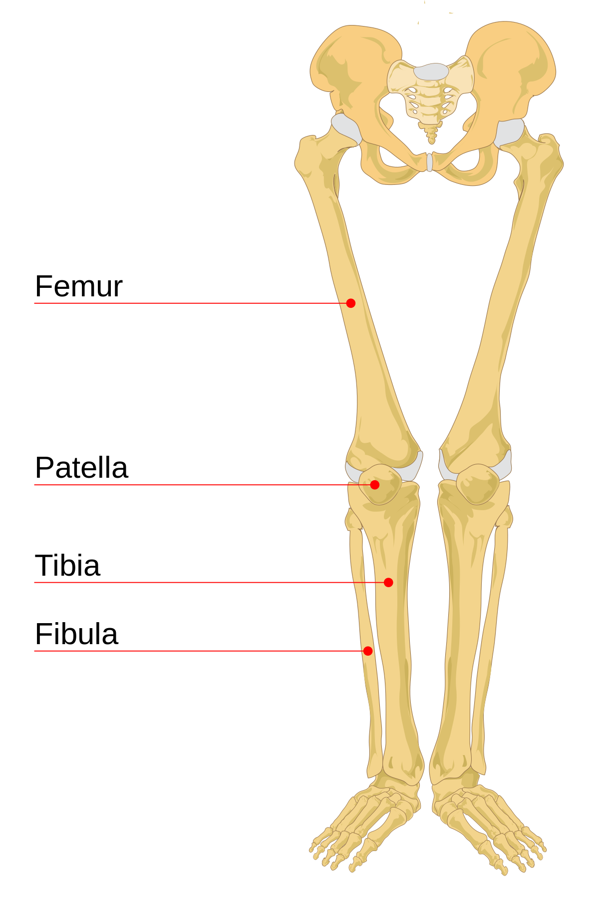
Leg bones diagram femur manual e books. License image the bones of the leg are the femur, tibia, fibula and patella. The foot bones shown in this diagram are the talus, navicular, cuneiform, cuboid, metatarsals. Muscles that lift the arches of the feet. Health diagram bone skeleton leg knee science anchor chart human human body.
Leg Bone Wikipedia from upload.wikimedia.org License image the bones of the leg are the femur, tibia, fibula and patella. The largest and most medial leg bone, forming both the knee and ankle joints. Muscles that lift the arches of the feet. Click now to learn more about the bones, muscles, and soft tissues tibia: The foot bones shown in this diagram are the talus, navicular, cuneiform, cuboid, metatarsals. The bones of the leg are the femur, tibia, fibula and patella. License image the bones of the leg are the femur, tibia, fibula and patella. The femur, or thighbone, is the longest and largest bone in the human body.
Download leg bone stock vectors.
Basic bone diagram enthusiast wiring diagrams. These bones are arranged into two major divisions: The bones of the leg are the femur, tibia, fibula and patella. Vector illustration with human skeleton scheme isolated on a white background. Ankle and foot pain massage therapy foot treatment. Bones of the leg and foot, lower leg bone anatomy, leg bones anatomy, leg muscles, leg bones diagram, leg bone structure, leg anatomy muscles, parts of the lower leg. The human leg, in the general word sense, is the entire lower limb of the human body, including the foot, thigh and even the hip or gluteal region. Your legs are two of your most important body parts. Long bones lengthen substantially as a person grows, and have a growth plate or epiphyseal plate at their ends, where new bone is formed during growth. Download leg bone stock vectors. Most relevant best selling latest uploads. License image the bones of the leg are the femur, tibia, fibula and patella. Skeleton leg ankle joints and toe phalanges, cuboid, metatarsal, navicular and cuneiform bones, hand drawn dorsal view of foot.
Reader view compact bone spongy bone of the long arm and leg bones that makes new red blood cells Long bones lengthen substantially as a person grows, and have a growth plate or epiphyseal plate at their ends, where new bone is formed during growth. They allow you to move and provide support for your upper body. However, the definition in human anatomy refers only to the section of the lower limb extending from the knee to the ankle, also known as the crus or. Affordable and search from millions of royalty free images, photos and vectors.
Tibia Leg Bone from www.ivyroses.com Learn vocabulary, terms and more with flashcards, games and other study tools. Looking for simple bone diagram barca fontanacountryinn com? Human foot bones anatomy sketch of orthopedics medicine. However, the definition in human anatomy refers only to the section of the lower limb extending from the knee to. Bone diagram barca fontanacountryinn com. Human bone diagram wiring diagrams click. The humerus and the femur are corresponding bones of the arms and legs, respectively. The red bone marrow inside of bones produces most of the blood cells, including erythrocytes (red blood cells), leukocytes (white blood cells), and thrombocytes (platelets).
Visit kenhub for more skeletal system quizzes.
Affordable and search from millions of royalty free images, photos and vectors. The largest and most medial leg bone, forming both the knee and ankle joints. Its lower end helps create the knee joint. Muscles that lift the arches of the feet. The radius and ulna (bones of the forearm), shown in supination (the arm rotated outward so that the palm. However, the definition in human anatomy refers only to the section of the lower limb extending from the knee to. Download leg bone stock vectors. Skeleton leg ankle joints and toe phalanges, cuboid, metatarsal, navicular and cuneiform bones, hand drawn dorsal view of foot. Learn vocabulary, terms and more with flashcards, games and other study tools. Your legs are two of your most important body parts. Learn how to draw the femur, patella, tibia, and fibula in this lesson! The femur, or thighbone, is the longest and largest bone in the human body. The bones of the leg are the femur, tibia, fibula and patella.
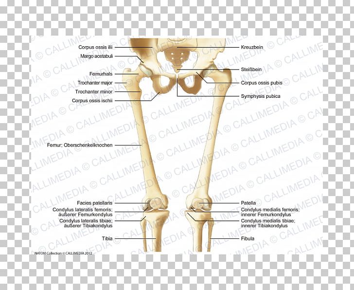
Pelvis definition, anatomy, diagram, & facts. Health diagram bone skeleton leg knee science anchor chart human human body. Leg femur diagram data wiring diagram today. The foot bones shown in this diagram are the talus, navicular, cuneiform, cuboid, metatarsals and calcaneus. Its lower end helps create the knee joint.
Finger Bone Hip Knee Human Leg Png Clipart Anatomy Angle Arm Bone Diagram Free Png Download from cdn.imgbin.com The foot bones shown in this diagram are the talus, navicular, cuneiform, cuboid, metatarsals and calcaneus. Skeleton leg ankle joints and toe phalanges, cuboid, metatarsal, navicular and cuneiform bones, hand drawn dorsal view of foot. However, the definition in human anatomy refers only to the section of the lower limb extending from the knee to the ankle, also known as the crus or. Click now to learn more about the bones, muscles, and soft tissues tibia: The foot bones shown in this diagram are the talus, navicular, cuneiform, cuboid, metatarsals. Ankle and foot pain massage therapy foot treatment. Looking for simple bone diagram barca fontanacountryinn com? Your legs are two of your most important body parts.
Download leg bone stock vectors.
Time to jump right into the biggest and strongest bones in the human body. Learn vocabulary, terms and more with flashcards, games and other study tools. Learn how to draw the femur, patella, tibia, and fibula in this lesson! They allow you to move and provide support for your upper body. Health diagram bone skeleton leg knee science anchor chart human human body. Muscles that lift the arches of the feet. The red bone marrow inside of bones produces most of the blood cells, including erythrocytes (red blood cells), leukocytes (white blood cells), and thrombocytes (platelets). The axial skeleton and the appendicular formed by the left and right hip bones, the pelvic girdle connects the lower limb (leg) bones to the axial. In children, most blood cells are produced by. Basic bone diagram enthusiast wiring diagrams. Its lower end helps create the knee joint. Reader view compact bone spongy bone of the long arm and leg bones that makes new red blood cells Master leg and knee anatomy using our topic page.


0 Comments